The 30th Annual NESM Symposium was held at the Marine Biological Laboratory in Woods Hole, MA on May 3rd, 2013, following a day of workshops. Warm spring sun and sea breezes welcomed the attendees and presenters to the Swope Center, nestled against picturesque Eel Pond in the center of MBL’s waterfront campus. During registration, a host of vendors from a wide range of companies set up display tables and discussed new developments in microscopy applications and burgeoning technology. The informal opening of the meeting gave attendees a chance to socialize over coffee and pastries.
Dr. Fettah Kosar, the President of the NESM board for 2013 opened the Symposium in the Swope Center’s spacious Meigs Room. Fettah gave an overview of NESM’s mission, and touched on how the society’s goals are served by meetings and presentations. Fettah also a brief history of the association between NESM and the MBL. Following Fettah’s introduction, Dr. Dejan Zecevic from the Yale University Medical School gave the first presentation of the day. He spoke about his research on dendritic spines and his use of voltage-sensitive dyes with low intensity fluorescence optical microscopy. Dr. Zecevic described the development of his research, partially inspired by his own experience as a former associate at the MBL. Dr. Zecevic’s work investigated the electrical function of fine structures called spines associated with dendritic fibers in neurons through a combination of advances in relatively inexpensive light sources, refinements of fine micro-scale voltage probes, and novel new dye chemistry. The combination of analytical techniques promises to aid in our understanding of the spines’ function, which is still nebulous.
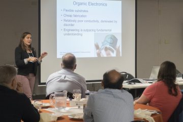
Dr. Thomas Tague of Bruker Optics, Inc. presented the next session, in which he detailed some novel applications of vibrational spectroscopy to the field of art history Dr. Tague presented an overview of the basic theory of vibrational spectroscopy, with a focus on Raman spectroscopy. He gave two examples of how Raman spectroscopy can be used to analyze art materials. The first application took advantage of Raman’s extremely fine spatial resolution to examine pigment and binder materials. Dr. Tague’s group used this sensitive technique to verify the authenticity of the Salvador Mundi, a lost Leonardo DaVinci painting. A significant amount of data about the origin of a work can be obtained through the combination of extensive optical documentation and per-phase chemical identification. Most importantly, this is accomplished with minimal detriment to the painting. The second example he gave championed a similar emphasis on preservation. Dr. Tague presented images of an on-site investigation using specially-designed Raman microscopes used to identify and catalog the pigments in cave paintings in sacred Native American sites in Texas. The technique was used to analyze both the original pigments on the cave paintings and the graffiti which has been slowly accumulating on the cave wall since the first settlers arrived. Because Raman spectroscopy is able to identify pigment particles without disturbing the rock surface, researchers were able to compare original and unwanted materials in order to design a laser ablation program that could effectively clean away defacing marks without damaging the underlying painting.
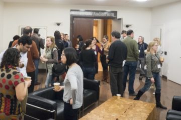
After the first two morning lectures, attendees visited an exhibiting vendor fair with a concurrent poster session from several graduate research students. Representatives were present from nearly 20 companies, ranging from software analysis manufacturers to microscope producers to supply vendors, and they were all ready to engage attendees in conversation and introduce them to new innovations for their research. Meanwhile, the students enthusiastically presented their work from a variety of fields. Poster topics included histological examination, fluorescence and marker examination of protein distribution, and correlative microscopy of microfibrils in cell structures. While all of the presentations were excellent, first prize went to Justine Allen, a student in the Brown University/MBL joint graduate program, who works in Dr. Roger Hanlon’s laboratory at the MBL. Her fascinating work on cuttlefish chromatophores was deemed to be the most in keeping with the spirit of versatility inherent in modern microscopy, and therefore narrowly clinched the top prize from the competition.
After lunch, the Symposium moved from Swope to the stately, amphitheater-style Lillie Auditorium. There, Dr. Kent McDonald from the University of California at Berkeley delivered the keynote lecture. Dr. McDonald’s talk expanded upon the workshop he gave the previous day on novel methods for rapidly fixing and preparing samples for transmission electron microscopy (TEM). Traditional TEM preparation protocols typically require very long staining, dehydration, and infiltration steps, and involve embedding samples in special epoxy or lowicryl resin which often takes days to polymerize. Dr. McDonald’s approach uses a carefully optimized system to greatly reduce preparation time from several days to just six to eight hours. His method uses cryogenic cooling, agitation, and special containers to increase the kinetics of water replacement and resin infiltration within the sample. Dr. McDonald showed that, due to these simple innovations, it is possible to prepare TEM-ready thin sections from fresh tissue in less than one day. The structure and tissue preservation is at least as good, and in some cases better, than what traditional preparation methods can provide. Dr. McDonald showed how this new methodology could streamline cumbersome sample prep– allowing electron microscopists to spend time on science, rather than on waiting for resin to polymerize.
After a brief coffee break sponsored by XRadia, Dr. Amy Griffin of the University of Delaware spoke about some in vivo neurophysiological techniques that allow for the activation and inactivation of discrete brain regions. She emphasized that these assays can be used to probe the functional interactions between different portions of the brain. Finally, Robert Brandom from Bruker Optics, Inc. gave the final talk of the afternoon, in which he described the use of energy-dispersive x-ray spectroscopy and electron backscatter diffraction in the SEM to analyze levels of corrosion in pipes at power plants. The 30th Annual Symposium was notable for the vast diversity of backgrounds from which its speakers came; from neuroscience to electron microscopy to power plant safety to art history, the day emphasized that microscopists are a very versatile group.
Jared Kelly & Rylie Walsh
2014 Clerk & 2014 Corresponding Secretary