This year NESM hosted two workshops at the 30th Annual Spring Symposium in Woods Hole. Kent McDonald from the Electron Microscopy Lab at UC Berkeley instructed attendees on high pressure freezing (HPF) and quick-freeze substitution (QFS), methods that he and Richard Webb (University of Queensland) co-developed for transmission electron microscopy. HPF enables high quality tissue preservation for biological samples that are thinner than 200 µm with out the need for chemical fixation. Historically, the process of embedding frozen samples into polymerized resin blocks has been a tedious one that takes several days. However, the QFS method that Kent demonstrated in the workshop requires only 6 hours to go from fresh tissue to blocks, ready to section. I have begun to implement these two techniques and so far they have worked nicely.
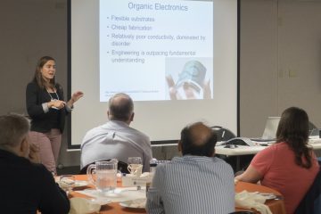
The second workshop was given by Thomas Tague Jr. (Bruker Optics, Inc) and focused on the uses of Fourier Transform Infrared Spectrocopy (FTIR) for both materials and biological sciences. This technique captures unique molecular fingerprints in the Mid-IR and has been especially useful for forensic investigations. Interestingly, FTIR in the mid-IR spectral band can identify specific molecule constituents which account for as low as <0. 1% of the sample. To demonstrate this, Thomas brought along the new, fully automated, LUMOS from Bruker.
George Bell
Director of Biological Sciences